Cardiology
At The Trowbridge Health Centre, we run a community cardiology service for the area.
This allows us to perform investigations that would normally be done in a hospital setting and is therefore convenient for the patient who does not then need to travel to a large district general hospital.
Dr Bimbh and his team perform echocardiograms and analyse 24 hour tapes. This is a service that we provide for NHS patients in the whole locality and access is through referral from your G.P.
A report of the tests is sent back to your own GP for any treatment necessary.
This service can only be accessed via a referral from your GP.
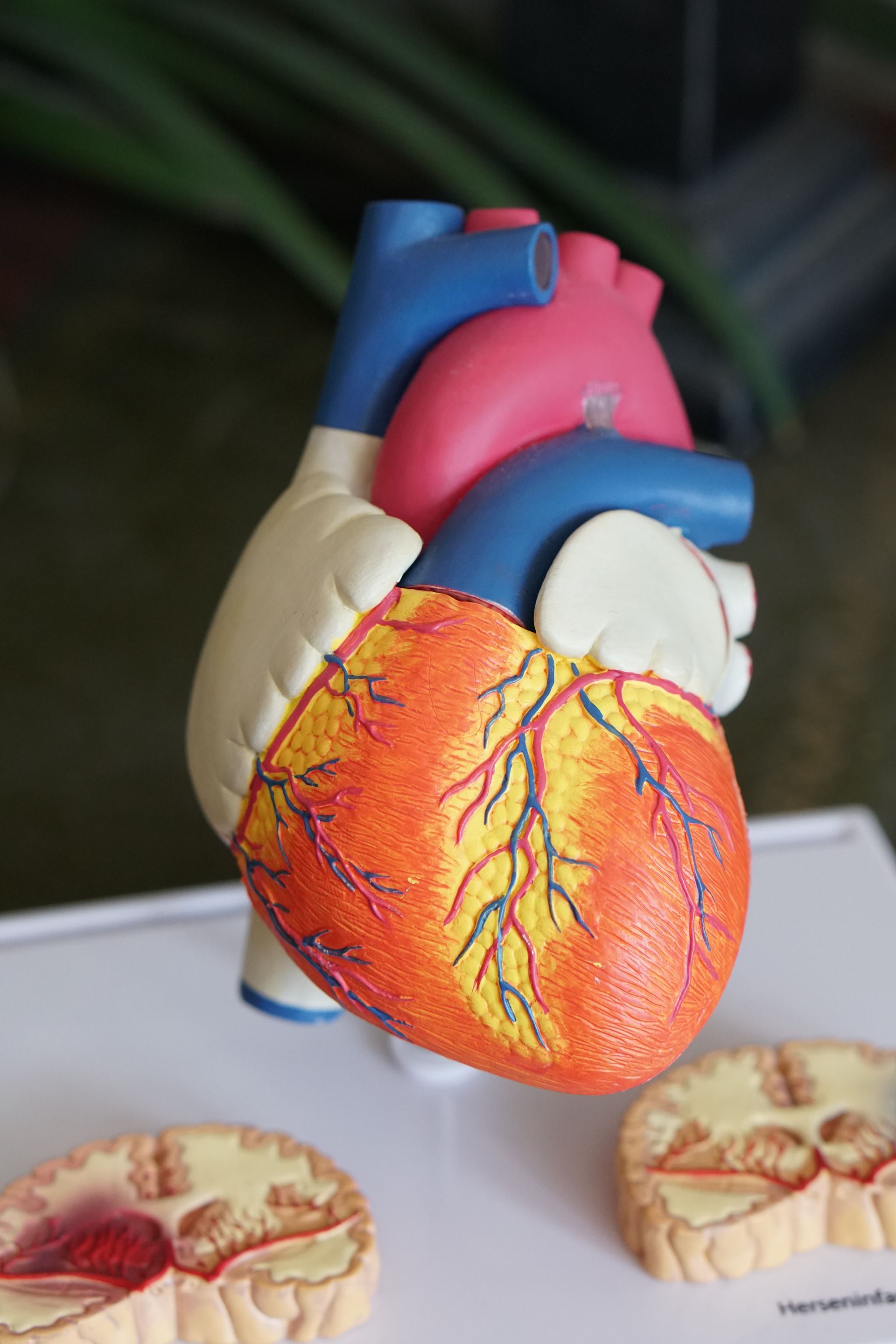
Echocardiogram
What is an echocardiogram?
An echocardiogram is an ultrasound scan of the heart. It is sometimes just called an 'ECHO'. Ultrasound is a very high frequency sound that you cannot hear, but it can be emitted and detected by special machines. The scan can give accurate pictures of the heart muscle, the heart chambers, and structures within the heart such as the valves.
What happens during the test?
You will need to undress to the waist and lie on the couch. A probe is placed on your chest (it is a bit like a very thick blunt pen). Also, lubricating jelly is put on the probe to ensure good contact with the skin. The probe is connected by a wire to the ultrasound machine and monitor. Pulses of ultrasound are sent from the probe through the skin towards your heart. The ultrasound waves then echo ('bounce back') from the heart and various structures in the heart.
The amount of ultrasound that echoes back depends on the density of the tissue the sound has hit. Therefore, the different structures send back different echoes. For example, ultrasound travels freely through fluid so there is little echo from blood in heart chambers. But, heart valves are dense tissues so ultrasound waves hitting a valve will echo back clearly.
The echoes are detected by the probe and are sent to the echocardiogram machine. They are displayed as a picture on the monitor. The picture is constantly updated so the scan can show movement as well as structure. (For example, the valves of a heart opening and closing.) The operator moves the probe around over the skin surface to get views from different angles. Some abnormalities can be seen quite clearly. For example, damaged heart valves, thickened heart muscle, some congenital heart defects, etc.
The test is painless and takes about 10-20 minutes. You may have to turn on your side during the test so the operator can scan the heart from different angles. You do not need any special preparation before the test. You eat and drink normally before and after the test. Continue to take your usual medication.
Ambulatory Electrocardiogram
What is an ambulatory electrocardiogram?
This test records the electrical activity of your heart when you are walking about (ambulatory) and doing your normal activities. Small metal electrodes are stuck onto your chest. Wires from the electrodes are connected to a small lightweight recorder (often called a Holter monitor). The recorder is attached to a belt which you wear round your waist. (It is like wearing a personal CD stereo.) The electrical activity is usually recorded for 24 hours.
Why is an ambulatory electrocardiogram test done?
Your doctor may advise this test if he or she suspects that you are having bouts of an abnormal heart rate or rhythm (arrhythmia). For example, if you have palpitations or episodes of dizziness. Some arrhythmias 'come and go', and may only last seconds or minutes. They may never be found when you are examined by a doctor. So, the test may detect an arrhythmia.
How is the test done?
It takes about 10 minutes for the electrodes and recorder to be fitted. You then go and do what you normally do over the next 24 hours. You wear the recorder when asleep in bed too. (However, you should not have a bath or shower as the recorder should not get wet.)
The ECG tracing is analysed at the end of the test. But, any times you record when you had symptoms will be most carefully analysed to see if you had an arrhythmia to account for the symptoms.
The monitor and electrodes are removed in another appointment approximately 24 hours after they were fitted. The ECG tracing is analysed and a report sent to your GP.
Page created: 10 March 2022